Facial moles are a common condition found in many people. They can be present at birth or develop over the course of a lifetime. Though they are frequently referred to as beauty marks, in many cases they are anything but beautiful. Non-verbal communication is key to how we communicate with the outside world, and the non-verbal communication messages are generally sent by our faces. A healthy facial complexion that is clear, bright and glowing sends a message of health and vitality. When we have pigment spots, bumps, or moles on our face, they can make us appear to be older or less healthy and vibrant.
Moles colors run the entire spectrum of browns, but occasionally may be black or blue. Facial moles are often benign and are not generally considered dangerous.
Moles exist both above and below the skin surface. The part of the mole that we see is often just the “tip of the iceberg”.
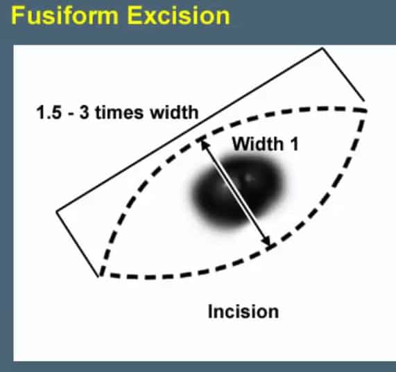
 Since moles extend so deeply beyond the top skin layer, the only way to fully remove them is through surgical excision. This is necessary when the mole has irregular cells or is cancerous. Fortunately, most moles are not dangerous but are instead a cosmetic concern. Because surgical excision leaves a scar that is often 3 times as long as the lesion itself, many people chose not to undergo surgical excision and instead continue to live with their irregular facial feature.
Thankfully, there is another option. This can only be used if the lesion isn’t comprised of irregular or cancerous cells and as such, requires a small portion of the mole to be removed and sent to a laboratory where is can be analyzed.
After injecting anesthesia, a small piece of the top layer of the lesion is shaved off and sent to the pathologist for evaluation.
The one problem with this method involves the “shaving process”. Using a blade, its often difficult to remove the lesion in a way that is precisely level with the surrounding tissue. Also at times, the biopsy can be too deep resulting in a “divot”.
The Hybrid Laser Mole Treatment involves:
- Shaving off a very small piece of the lesion to ensure that it is benign and doesn’t require a wider excision.
- Waiting for the pathology report to confirm that the lesion is benign.
- When the benign character of the lesion is confirmed, the portion of the lesion that remains is treated using a fine 2 mm spot size Erbium Laser. The lesion is planed down cell layer-by-cell layer until it is level with the surrounding skin.
- The wound is covered with a healing polymer called Kericure and the patient is instructed to keep it moist with a healing ointment.
- The body will heal the wound over a one-to-two week period, and what will be left behind is a flat contour where the mole previously existed.
It should be noted that this procedure does leave the deeper portion of the lesion behind, but since they generally grow very slowly, the results are likely to last for a very long time.
This procedure is a great option for use on the nose, where a divot from a traditional shave biopsy would be very noticeable. It also works well on other complicated parts of the face.
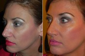
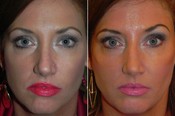
Here is a picture of a patient who is a fashion model and who had a small mole on her nose. This detracted from her facial appearance. A mole in this central portion of the face can be very difficult to remove without creating an irregular contour.
By using my Hybrid Laser Mole Treatment I was able to finesse her mole back to a flatter, more attractive and more harmonious appearance.
Pre and post Hybrid Laser Mole Treatment
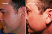
This gentleman’s mole was in his sideburn area. He was frustrated by his inability to shave, so I first took a small biopsy to ensure it was benign. I then used a 2 mm spot size Erbium Laser to plane down the lesion layer by layer until it was level with the surrounding skin. He was very pleased with the appearance of his skin after he healed, and now he doesn’t need to wear his sideburns as long as he did before.


